Chapter 2
Cell Structure

Courtesy of Tutor Vista
Courtesy of Buzzle.com
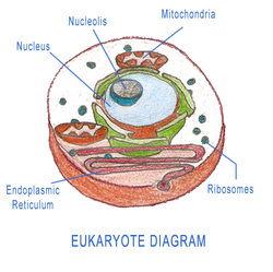
Cells are the smallest unit that is capable of performing life functions. Cells come in many shapes and sizes. The two types of cells are prokaryotic and ekaryotic. Prokaryotic cells are found in one-celled organisms and do not have membrane-bound structures. Bacteria is an example of a prokaryotic cell. Eukaryotic cells have membrane-bound structures. Protists, fungi, plants, and animals are examples of eukaryotic cells.
Parts of a Cell
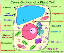
Cell walls are tough, rigid outer coverings that protect the cell and give it shape. Plants, algae, fungi, and most bacteria have cell walls. Animal cells do not have cell walls.
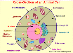
Cell membranes provide a protective layer around all cells and regulate interactions between the cell and the environment. If a cell has a cell wall, the cell membrane is just inside of it.
Cytoplasm is a gelatinlike substance that constantly flows inside the cell membrane. The cytoskeleton, which helps a cell maintian or change its shape, is found in the cytoplasm. Organelles of eukaryotic cells are also found in the cytoplasm. Organelles help a cell carry on life processes and are ususally separated from the cytoplasm by membranes.
The nucleus directs all activities of the cell. It is usually the largest organelle in a cell and contains the hereditary material for the cell. A nucleolus, responsible for making ribosomes, is found in the nucleus.
Chloroplasts are green organelles found in plant cells. They capture light energy and use it to make sugar that the plant cell uses for food. Animal cells do not have chloroplasts for making food. They must get food from the environment.
Mitochondria are organelles that release energy from food. More active cells, like muscle cells, have more mitochondria because they need more energy.
Ribosomes are organelles that are not membrane bound. They are the site where proteins are made. Proteins are important to a cell because they take part in nearly every cell activity. Some ribosomes float freely in the cytoplasm, and are known as free ribosomes, and other ribosomes are attached to the endoplasmic reticulum, and are known as bound ribosomes.
Endoplasmic reticulum, or ER, is an organelle that extends from the nucleus to the cell membrane. It is a series of folded membranes in which materials can be processed and moved inside or outside of the cell. ER with no ribosomes attached is called smooth endoplasmic reticulum. ER with attached ribosomes is called rough endoplasmic reticulum.
Golgi Bodies, or Golgi Apparatus, are organelles that sort proteins and other substances in the cell and package them into membrane bound structures called vesicles. These vesicles carry cellular substances to other places in the cell. They also carry substances to the cell membrane so that they can be released from the cell to go to another cell.
Vacuoles are membrane bound organelles that can store almost anything the cell needs it to. In a plant cell, the vacuole takes up most of the space. In an animal cell, the vacuole is surprisingly small.
Lysosomes are membrane bound organelles that break down food, wastes, and worn out cell parts. Lysosomes contain acidic enzymes. If there wasn't a membrane around the lysosome, the enzymes would leak into the cell and destroy it.
Courtesy of Enchanted Learning
Courtesy of Enchanted Learning
While many one-celled organisms can carry on all their life functions by themselves, many-celled organisms cannot. In many-celled organisms, cells are organized into tissues. Tissues are groups of similar cells that work together to do one job. Tissues are organized into organs. Organs are structures made up of two or more different types of tissues that work together. Organs are grouped into organ systems that work together to perform certain functions.
Viewing Cells

Courtesy of Qwick Step
The first microscope was made by a Dutch maker of reading glasses in the late 1500s. He put two lenses together in a tube and got a larger image than one of the lenses could make by itself. In the mid 1600s, Antonie van Leeuwenhoek made a microscope with a small glass bead as the lens. This microscope could magnify up to 270 times! Using this, he saw things in pond water that people could never see before.
Depending on the number of lenses a microscope has, it is given a different name. A simple microscope only has one lens. Another kind of microscope is a compound light microscope. It has two sets of lenses: eyepiece lenses and objective lenses. To determine the magnifications of a compound microscope, multiply the power of the objective lens and the power of the eyepiece lens. Microscopes with two eyepieces are called stereomicroscopes and allow you to see three-dimensional images. Electron microscopes use magnetic fields to direct beams of electrons and are used to view images that can't be seen with other microscopes. Some electron microscopes can magnify images up to one million times!
In 1665, Robert Hooke looked at a piece of cork under a microscope. It looked like the cork was made up of empty little boxes. Hooke named these boxes cells. In the 1830s, Matthias Schlieden studied plants under microscopes. He concluded that all plants are made up of cells. Theodor Schwann studied animal cells and concluded that all animals are made up of cells. Later, they put their ideas together and concluded that all living things are made up of cells. Many years later, Rudolph Virchow proposed that cells divide to form new cells and that every cell came from a cell that already existed. His work and the work of many other scientists developed into the cell theory. The cell theory states that all organisms are made up of one or more cells, the cell is the basic unit of structure and function in organisms, and all cells come from cells.
Viruses

Courtesy of leavingbio.net
A virus is a strand of hereditary material surrounded by a protein coating. Viruses can only make copies of themselves, and they need the help of living host cells to do this. First a virus attaches to a living host cell. Then the virus shoots its DNA into the cell. The viral DNA instructs the host cell to make more viral DNA and parts. The living host cell does what the viral DNA tells it to do and makes new viruses. The host cell finally bursts open, releasing the new viruses, and is destroyed.
A virus can either be active or latent. An active virus is alive and destroying living host cells. A latent virus is also alive but not destroying living host cells. A latent virus can become active at any time.
Viruses can attack all living things, but most viruses can only attack one kind of living host cell. This is because a virus must attach to a living host cell exactly. A virus cannot move on its own. It can be moved by the wind or inhaled.
There aren't many ways that viruses can be treated. Antibiotics can only treat bacterial infections and don't work to stop viral diseases. Your body can produce interferons to to protect non-infected cells. There are some antiviral drugs that can help fight viruses, but they aren't used very often because of their adverse side effects. Viruses can be prevented by vaccinating people, improving sanitary conditions, quarantining patients, and controlling animals that spread disease. Vaccines are weakened or dead viruses that are injected into your body so that you can become immune to that certain virus.
Viruses aren't always harmful. One good use of a virus is gene therapy. This is when normal hereditary material enclosed in viruses enters cells with defective hereditary material and replaces it with the new, normal hereditary material. Someday, gene therapy might be used to cure cancer.
